

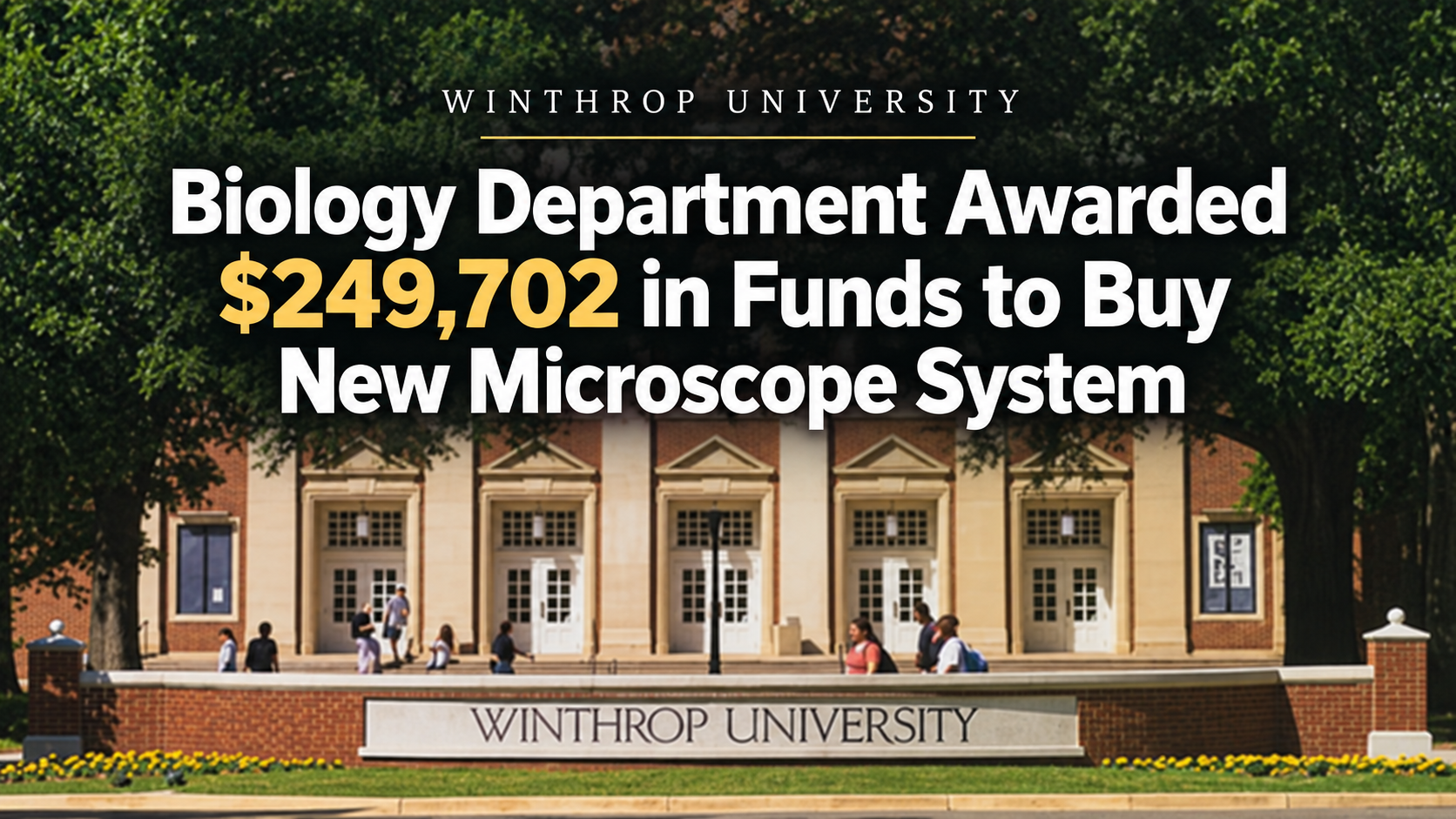
 ROCK HILL, SOUTH CAROLINA – Winthrop University’s Department of Biology was awarded a $249,702 federal grant this summer to purchase a sophisticated microscope system to enhance its biomedical research and to replace aging equipment.
The equipment, called a Spinning Disk Echo Confocal Microscope, will strengthen basic research, such as cancer therapy and vision research, according to Assistant Professor Jena Chojnowski. Essential for the progress and success of faculty research, the equipment will aid in student research, student training in classes, recruitment of new faculty and outreach programs at the university.
The grant is awarded through the National Institute of General Medical Sciences, a division of the National Institutes of Health.
A confocal microscope is an advanced type of light microscope that uses a laser and a pinhole aperture to block out-of-focus light, producing sharp, high-resolution 3D images. It takes multiple optical slices at different depths and uses a computer to build a detailed 3D model.
Chojnowski said the microscope system will be installed in Dalton Hall’s microscopy facility sometime in the fall. It will modernize undergraduate and graduate training in the life sciences program through course work and experiential learning that will allow future generations to experience a piece of modern technology that drives biomedical advancements.
“Research and education across departments, nine research labs, and at least four classes will be impacted by the cutting-edge yet easy-to-use spinning disk technology of the Echo microscope,” said Chojnowski, adding that students in the Department of Biology and Department of Chemistry, Physics, Geology, and the Environment will benefit from the equipment.
Two Main Components
The high-resolution unit contains two main components: an inverted Revolution microscope that includes a computer and cameras and the spinning disk confocal laser unit. The automated and inverted multi-dimensional imaging unit allows for multi-point, mosaic, and multi-channel Z-stack imaging capabilities.
It will provide the opportunity to acquire high-resolution 3D immunofluorescence images through an easy-to-use interface for experienced microscopists and non-microscopy users alike. The Echo microscope will be equipped with six Apo or fluorite phase objectives (2x, 4x, 10x, 20x, 40x, and a 60x oil), four laser lines (405, 488, 555, and 640), and two cameras (one monochrome 95% QE with 2048×2048 and one color 5MP with 2448×2048) that will meet the needs of all users.
Currently, faculty and students are relying on four fluorescent microscopes (non-confocal) purchased before 2008 or outsourcing to other universities for their imaging. The university’s 17-year-old Olympus FV-1000 spectral laser-scanning confocal microscope has recently fully malfunctioned due to mechanical failure of the laser throughput and due to its advanced age, the mechanical parts are no longer in production and are unavailable for purchase.
Chojnowski said the lack of a functional confocal microscope is hindering the departments’ ability to perform top-tier research without external support, which is financially prohibitive for some of the university’s researchers, and limits their ability to teach specific classes such as BIOL530, Current Methods in Microscopy. Therefore, the biology and other departments had a significant need for a new, fully functional confocal imaging system like the Echo microscope that allows for diverse imaging applications, including Z-stacking, image stitching of large tissue sections, and fast acquisition of 3D volumetric multi-channel images.
These features will be used for a diverse range of NIH and SC INBRE funded biomedical research projects such as cellular transcriptional regulation of glioblastomas and prostate cancer, fluorescent tagging of neurotransmitters, corneal and retinal development and maintenance, and tissue-specific DNA repair mutants in Drosophila.
The purchase of this equipment was made through the Instrumentation Program for Resource Limited Institutions (RLI-S10, PAR-27-067) through the National Institute of General Medical Sciences of the National Institutes of Health under Award Number S10GM164748.
For more information, contact Chojnowski at [email protected].

Sign up for our Sunday Spectator. Delivered to your inbox every Sunday, with all the news from the week.

Thomas Hyslip lives in Tega Cay with his wife and daughter. After 27 years in the U.S. Army and Federal Law Enforcement, he retired to pursue his passion for teaching. Tom is now an Assistant Professor of Instruction at the University of South Florida. In 2 short years he has won 10 awards from the South Carolina Press Association, including first place in column writing, education beat reporting and best podcast.